Torzija testikularnih apendiksa
| Torzija testikularnih apendiksa | |
|---|---|
 | |
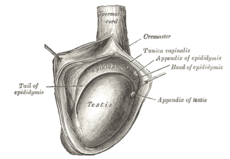
| Sa desne strane testisa i epididimisa, kroz otvorenu tuniki vaginalis, vidi se apndiksi. | |
| Specijalnost | Urologija |
| Klasifikacija i eksterni resursi | |
| ICD-10 | N44 |
| ICD-9 | 608.2 |
| OMIM | 187400 |
| DiseasesDB | 12984 |
| MedlinePlus | 000517 |
| eMedicine | med/2780 |
| MeSH | D013086 |
Torzija testikularnih apendiksa ili Morgagnijeva hidatida je je uvrtanje, oko svoje peteljke, malih produžetaka-apendiksa u obliku polipa (pedunkuliranih ili sesilnih). Najčešći je uzrok akutnog skrotuma dečaka u prepubertetu.[1] Za razliku od torzije testisa, stanje nije hitno u smislu opstanka testikularnog tkiva.[2]
Anatomija[uredi | uredi kod]
Apendiksi skrotuma, ili mali produžeci u vidu polipa, su ostaci Milerovih ili Volfovih struktura:[3]
- Milerove strukture su ostaci apendiksa u području gornjeg pola testisa
- Volfove strukture su ostaci apendiksa u području epididimisa (pasemenika).
Apendiks testisa prisutn je u 92% svih testisa i obično se nalazi u području gornjeg pola testisa u udubljenje između testisa i pasemenika. Apendiks pasemenik prisutn je u 23% testisa i obično projektuje iz glave pasemenika (epididimisa), mada njegova lokacija može da varira.
Etiopatogeneza[uredi | uredi kod]
Torzija testikularnih apendiksa nastaje uvrtanjem, oko svoje peteljke, malih produžetaka u obliku polipa (pedunkuliranih ili sesilnih) koji se mogu javiti na gornjim polovima epididimisa i testis. Njihovim uvrtanjem nastaje prekid cirkulacije. Tada nastaje klinička slika koja je skoro istovetna sa kliničkom slikom torzije semene vrpce
Klinička slika[uredi | uredi kod]
Klinička slika karakteriše se bolom, najčešće manjeg intenziteta nego kod torzije testisa. Deca se javljaju nakon više od jednog dana tegoba jer se bolovi javljaju nešto sporiji.
Dijagnoza[uredi | uredi kod]
Karakteristična je bolnost pri palpaciji kvržice iznad gornjeg pola testisa i koja ima karakter torkvirana Morgagnijeva hidatide koja se prosijava plavičasto kroz kožu skrotuma.[4] Dijagnoza se uspešno postavlja primenom ultrasonografskog pregleda testisa.[5][6]
Odlaganjem dijagnoze može doći do gnojne upale koja daljim širenjem može izazvati epididimoorhitis.[7][8][9]
Diferencijalna dijagnoza[uredi | uredi kod]
Terapija[uredi | uredi kod]
Ne postoji jedinstveni stav oko terapije. Neki zagovaraju konzervativnu terapiju: pošteda, antibiotici i ekspplorativni pristup.[10]
Bol obično prestaje u roku od jedne nedelje, ali može trajati nekoliko nedelja. Za njegovo lečenje koriste se analgetici iz grupe NSAIL i obloge sa ledom.[11]
Hirurško lečenje preporučuje se zbog opasnosti od propuštanja torzije testisa. Zahvat je kratak, uključuje odstranjenje torkviranog apendiksa, a oporavak je kratak uz brzo vraćanje telesnim aktivnostima.
Stav većeg broja dečjih hirurga je da se svaki akutni skrotum eksplorira, pa tako i u slučaju dijagnoze torzije testikularnog priveska.[12]
Izvori[uredi | uredi kod]
- ↑ Nason GJ, Tareen F, McLoughlin D, McDowell D, Cianci F, Mortell A. Scrotal exploration for acute scrotal pain: a 10-year experience in two tertiary referral paediatric units. Scand J Urol. Oct 2013; 47 (5): 418-22.
- ↑ Sahni D, Jit I, Joshi K, Sanjeev. Incidence and structure of the appendices of the testis and epididymis. J Anat. 1996 Oct. 189 ( Pt 2):341-8. [Medline].
- ↑ Kogan SJ, Hadziselmovic F, Howards SS. Pediatric andrology: congenital and acquired scrotal abnormalities. Adult and Pediatric Urology. 4th ed. 2002. Vol 3: 2570-2581.
- ↑ * Barloon TJ, Weissman AM, Kahn D. Diagnostic imaging of patients with acute scrotal pain. Am Fam Physician. 1996 Apr. 53(5):1734-50.
- ↑ Strauss S, Faingold R, Manor H. Torsion of the testicular appendages: sonographic appearance. J Ultrasound Med. 1997 Mar. 16(3):189-92; quiz 193-4.
- ↑ Aydogdu O, Burgu B, Gocun PU, Ozden E, Yaman O, Soygur T, et al. Near infrared spectroscopy to diagnose experimental testicular torsion: comparison with Doppler ultrasound and immunohistochemical correlation of tissue oxygenation and viability. J Urol. 2012 Feb. 187(2):744-50.
- ↑ Boettcher M, Bergholz R, Krebs TF, WenkeK, Treszl A, Aronson DC et al. Differentiationof epididymitis and appendix testis torsion byclinical and ultrasound signs in children. Urology.Oct 2013; 82 (4): 899-904.
- ↑ Rakha E, Puls F, Saidul I, Furness P. Torsionof the testicular appendix: importance of associatedacute inflammation. J Clin Pathol. Aug2006; 59 (8): 831-4.
- ↑ Karmazyn B, Steinberg R, Livne P, KornreichL, Grozovski S, Schwarz M et al. Duplex sonographicfindings in children with torsion of thetesticular appendages: overlap with epididymitis and epididymoorchitis. J Pediatr Surg. Mar 2006; 41 (3): 500-4.
- ↑ Watkin NA, Reiger NA, Moisey CU. Is the conservative management of the acute scrotum justified on clinical grounds?. Br J Urol. 1996 Oct. 78(4):623-7.
- ↑ Holland JM, Graham JB, Ignatoff JM. Conservative management of twisted testicular appendages. J Urol. 1981 Feb. 125(2):213-4. [Medline].
- ↑ Even L, Abbo O, Le Mandat A, Lemasson F, Carfagna L, Soler P, et al. Testicular torsion in children: Factors influencing delayed treatment and orchiectomy rate. Arch Pediatr. 2013 Apr. 20(4):364-8.
Literatura[uredi | uredi kod]
- Nason GJ, Tareen F, McLoughlin D, McDowell D, Cianci F, Mortell A. Scrotal exploration for acute scrotal pain: a 10-year experience in two tertiary referral paediatric units. Scand J Urol. 2013 Oct. 47(5):418-22.
- Rakha E, Puls F, Saidul I, Furness P. Torsion of the testicular appendix: importance of associated acute inflammation. J Clin Pathol. 2006 Aug. 59(8):831-4.
- Boettcher M, Bergholz R, Krebs TF, Wenke K, Aronson DC. Clinical predictors of testicular torsion in children. Urology. 2012 Mar. 79(3):670-4.
- Karmazyn B, Steinberg R, Kornreich L. Clinical and sonographic criteria of acute scrotum in children: a retrospective study of 172 boys. Pediatr Radiol. 2005 Mar. 35(3):302-10.
- Boettcher M, Bergholz R, Krebs TF, Wenke K, Treszl A, Aronson DC, et al. Differentiation of epididymitis and appendix testis torsion by clinical and ultrasound signs in children. Urology. 2013 Oct. 82(4):899-904.
- Pepe P, Panella P, Pennisi M, Aragona F. Does color Doppler sonography improve the clinical assessment of patients with acute scrotum?. Eur J Radiol. 2006 Oct. 60(1):120-4.
- Melloul M, Paz A, Lask D, et al. The pattern of radionuclide scrotal scan in torsion of testicular appendages. Eur J Nucl Med. 1996 Aug. 23(8):967-70.
- Saxena AK, Castellani C, Ruttenstock EM, Höllwarth ME. Testicular Torsion: A 15-Year Single Center Clinical and Histological Analysis. Acta Paediatr. 2012 Mar 3.
- Fisher R, Walker J. The acute paediatric scrotum. Br J Hosp Med. 1994 Mar 16-Apr 5. 51(6):290-2.
- Hormann M, Balassy C, Philipp MO, Pumberger W. Imaging of the scrotum in children. Eur Radiol. 2004 Jun. 14(6):974-83.
- Johnson KA, Dewbury KC. Ultrasound imaging of the appendix testis and appendix epididymis. Clin Radiol. 1996 May. 51(5):335-7.
- Kadish HA, Bolte RG. A retrospective review of pediatric patients with epididymitis, testicular torsion, and torsion of testicular appendages. Pediatrics. 1998 Jul. 102(1 Pt 1):73-6.
- Lewis AG, Bukowski TP, Jarvis PD, et al. Evaluation of acute scrotum in the emergency department. J Pediatr Surg. 1995 Feb. 30(2):277-81; discussion 281-2.
- McAndrew HF, Pemberton R, Kikiros CS. The incidence and investigation of acute scrotal problems in children. Peditric Surg Int. 2002 Sept. 18:435-437.
- Rabinowitz R, Hulbert WC Jr. Acute scrotal swelling. Urol Clin North Am. 1995 Feb. 22(1):101-5.
- Ravichandran S, Blades RA, Watson ME. Torsion of the epididymis: a rare cause of acute scrotum. Int J Urol. 2003 Oct. 10(10):556-7.
- Siegel MJ. The acute scrotum. Radiol Clin North Am. 1997 Jul. 35(4):959-76.
- Williamson RC. Torsion of the testis and allied conditions. Br J Surg. 1976 Jun. 63(6):465-76.
- Yazbeck S, Patriquin HB. Accuracy of Doppler sonography in the evaluation of acute conditions of the scrotum in children. J Pediatr Surg. 1994 Sep. 29(9):1270-2.