Datoteka:Cytokinesis-electron-micrograph.jpg
Cytokinesis-electron-micrograph.jpg (745 × 451 piksela, veličina datoteke/fajla: 200 kB, MIME tip: image/jpeg)
Picture from English Wikipedia
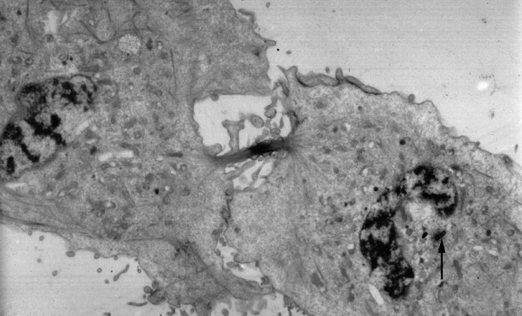
An electron micrograph image of a cell that has almost completed cell division and cytokinesis. Mitosis has already been completed. An arrow points to a centrosome still present near one of the nuclei.
From http://www.wadsworth.org/bms/SCBlinks/web_mit2/RES_MIT.htg/teleoph.jpg archive copy at the Wayback Machine, the Wadsworth Center, which is part of the New York State Department of Health and devoted to public education. Since it's part of the US government, I'll assume public domain.
This work is in the public domain in the United States because it is a work prepared by an officer or employee of the United States Government as part of that person’s official duties under the terms of Title 17, Chapter 1, Section 105 of the US Code.
Note: This only applies to original works of the Federal Government and not to the work of any individual U.S. state, territory, commonwealth, county, municipality, or any other subdivision. This template also does not apply to postage stamp designs published by the United States Postal Service since 1978. (See § 313.6(C)(1) of Compendium of U.S. Copyright Office Practices). It also does not apply to certain US coins; see The US Mint Terms of Use.
|
 | |
| Ova datoteka je identificirana kao slobodna od poznatih ograničenja po zakonu o autorskim pravima, uključujući sva povezana i srodna prava. | ||
Uploaded 07:21, 21 July 2005 .by user . Natalinasmpf . . 745x451 (87580 bytes) (An electron micrograph image of a cell that has almost completed cell division and cytokinesis. Mitosis has already been completed. An arrow points to a centrosome still present near one of the nuclei.
This work is in the public domain in the United States because it is a work prepared by an officer or employee of the United States Government as part of that person’s official duties under the terms of Title 17, Chapter 1, Section 105 of the US Code.
Note: This only applies to original works of the Federal Government and not to the work of any individual U.S. state, territory, commonwealth, county, municipality, or any other subdivision. This template also does not apply to postage stamp designs published by the United States Postal Service since 1978. (See § 313.6(C)(1) of Compendium of U.S. Copyright Office Practices). It also does not apply to certain US coins; see The US Mint Terms of Use.
|
| |
| Ova datoteka je identificirana kao slobodna od poznatih ograničenja po zakonu o autorskim pravima, uključujući sva povezana i srodna prava. | ||
)
Historija datoteke
Kliknite na datum/vrijeme da biste vidjeli tadašnju verziju datoteke.
| Datum/vrijeme | Minijatura | Dimenzije | Korisnik | Komentar | |
|---|---|---|---|---|---|
| aktualna | 14:54, 24 maj 2011 | | 745 × 451 (200 kB) | Zephyris | Reverted to version as of 12:52, 24 May 2011 |
| 14:54, 24 maj 2011 |  | 745 × 451 (200 kB) | Zephyris | Reverted to version as of 12:50, 24 May 2011 Reversion seemed not to work | |
| 14:52, 24 maj 2011 |  | 745 × 451 (200 kB) | Zephyris | Reverted to version as of 12:50, 24 May 2011 Confusion with cached images | |
| 14:52, 24 maj 2011 |  | 745 × 451 (200 kB) | Zephyris | Oops, uploaded the original file last time by accident! | |
| 14:50, 24 maj 2011 |  | 745 × 451 (200 kB) | Zephyris | Inverted image: It is more common to show more intensly absorbing features in an electron micrograph (e.g. chromatin and the midbody) as dark rather than light. Asjusted levels and contrast: To both use the full histogram range and emphasise detail in the | |
| 19:43, 1 decembar 2005 |  | 745 × 451 (86 kB) | Rasbak | Picture from English Wikipedia An electron micrograph image of a cell that has almost completed cell division and cytokinesis. Mitosis has already been completed. An arrow points to a centrosome still present near one of the nuclei. |
Upotreba datoteke
Sljedeća stranica koristi ovu datoteku:
Globalna upotreba datoteke
Ovu datoteku upotrebljavaju i sljedeći projekti:
- Upotreba na projektu ar.wikipedia.org
- Upotreba na projektu bn.wikipedia.org
- Upotreba na projektu ca.wikipedia.org
- Upotreba na projektu en.wikipedia.org
- Upotreba na projektu es.wikipedia.org
- Upotreba na projektu gl.wikipedia.org
- Upotreba na projektu ht.wikipedia.org
- Upotreba na projektu hy.wikipedia.org
- Upotreba na projektu it.wikipedia.org
- Upotreba na projektu ja.wikipedia.org
- Upotreba na projektu kk.wikipedia.org
- Upotreba na projektu nl.wikipedia.org
- Upotreba na projektu nl.wikibooks.org
- Upotreba na projektu pl.wikipedia.org
- Upotreba na projektu pt.wikipedia.org
- Upotreba na projektu ru.wikipedia.org
- Upotreba na projektu sl.wikipedia.org
- Upotreba na projektu sr.wikipedia.org
- Upotreba na projektu tr.wikipedia.org
- Upotreba na projektu uk.wikipedia.org